Currently Empty: £0.00
GCSE Biology: How Microscopes Changed Biology—From Blurry to Brilliant
April 23, 2025

Introduction
Think back to your first time using a microscope at school. You might have looked at a slice of onion or a drop of pond water. You saw something — but probably not in great detail. That’s because most school microscopes are light microscopes, which have limited magnification and resolution.
Now, imagine being able to zoom in so far that you can see the folds inside a mitochondrion or spot individual ribosomes. That’s the power of electron microscopes. They take blurry, vague shapes and reveal crisp, highly detailed structures. This improvement hasn’t just helped us see cells more clearly — it’s helped scientists understand them on a much deeper level.
Spec Point 1.3 — What You Need To Know
This topic falls under the GCSE Biology specification. You should be able to:
- Explain how microscope developments, especially electron microscopes, deepened our understanding of cells
- Compare light and electron microscopes
- Understand and use the concepts of magnification and resolution
- Apply the magnification formula: I = A × M (Image size = Actual size × Magnification)
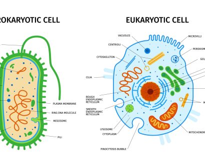
What Is a Light Microscope?
Light microscopes use visible light and glass lenses to magnify samples. They’re the standard in classrooms and labs, typically magnifying up to 2,000 times.
Advantages:
- Portable and easy to use
- Affordable – ideal for schools
- Can be used to observe living cells
Limitations:
- Lower magnification compared to electron microscopes
- Limited resolution — fine details are often blurred
What Is an Electron Microscope?
Electron microscopes use beams of electrons instead of light. These electrons interact with the specimen to form a detailed image on a screen. They can magnify up to 2,000,000 times.
Advantages:
- Far greater magnification and resolution
- Reveals fine cell structures and small organelles
Limitations:
- Can’t be used on living samples (requires a vacuum environment)
- Expensive and bulky
- Needs specialist training to operate and interpret results
Why This Matters in Biology
Before electron microscopes, much of cell biology was guesswork. Internal structures were hard to study, and tiny organelles remained hidden. Electron microscopes changed that. We’ve since discovered new organelles, observed viruses in detail, and tracked how diseases alter cells.
This tech has driven advances in genetics, medicine, vaccines, and more. So, this topic isn’t just for the exam — it’s about real-world science.
Quick Comparison Table
| Feature | Light Microscope | Electron Microscope |
|---|---|---|
| Magnification | Up to 2,000× | Up to 2,000,000× |
| Resolution | Lower | Much higher |
| Specimen Type | Living or non-living | Non-living only |
| Portability | Lightweight and portable | Large and stationary |
| Cost | Inexpensive | Expensive |
3-Mark Exam Strategy
When answering a question about why electron microscopes are better, remember to include these three points:
- Higher magnification
- Higher resolution
- More detailed images
That’s how you secure all 3 marks.
Key Terms to Know
- Magnification – How many times bigger the image is than the actual object
- Resolution – The ability to distinguish between two points that are close together
- Clarity – A non-technical term often used to describe image sharpness
Magnification Formula: I = A × M
Use this formula to calculate magnification in your exam:
Magnification = Image Size ÷ Actual Size
To remember it:
- I = Image size
- A = Actual size
- M = Magnification
Triangle method tip:
Draw a triangle with I on top and A and M underneath. Cover the one you’re solving for.
Worked Example:
Question: A real cell is 0.04 mm and appears 20 mm under the microscope. What’s the magnification?
Step 1: Use the formula: M = I ÷ A
Step 2: 20 ÷ 0.04 = 500
Answer: 500×
AMAL Top Tips for Students
- Practise calculations using the magnification formula — watch out for unit conversions
- Learn to label diagrams of light and electron microscopes
- Memorise the 3-mark answer points
- Understand and define resolution — it’s a frequent mark scheme keyword
FAQs
Q: Why can’t electron microscopes be used on live cells?
A: They require a vacuum and metal coatings, which destroy living tissue.
Q: What’s the main benefit of electron microscopes?
A: Their higher magnification and resolution let us see far more detail.
Q: Do I lose marks for missing units?
A: Yes. Always include the unit — like 500× — in your final answer.
Final Thought
If you understand how microscopes have advanced biology and you can confidently apply the I = A × M formula, you’re ready for this part of your GCSE. Electron microscopes transformed science — and now you know why.
More Support & Resources
🎥 YouTube Playlist:
📚 AMALathon Learning Series:
📝 AMALathon 2 Registration: